
Study Up For The CSCS! Chapter 1
Chapter 1: Structure and Function of the Muscular, Neuromuscular, Cardiovascular and Respiratory Systems
The systems of the body that produce movements create a network of interaction with each other that allow us to create purposeful, effective and coordinated movements for the activities of sport and daily life. These movements are the result of muscle fibers acting collectively to pull on our bones which act as levers, and generate force. Muscle fiber firing is controlled by our nervous system, driving electrical signals from the cerebral cortex down through the spine and along the efferent (outgoing) motor nerves, across the sarcolemma and into the muscle cells. And let’s not forget the importance of the cardiovascular system to deliver oxygen and remove waste to allow these processes to take place. All of these systems allow us to move and exert force, but there is much more detail than what is presented here, let’s look deeper at the muscular system and understand how these structures function.

Photo Source: http://www.moralesbiology.com/uploads/9/7/8/6/9786451/9421995_orig.jpg?463
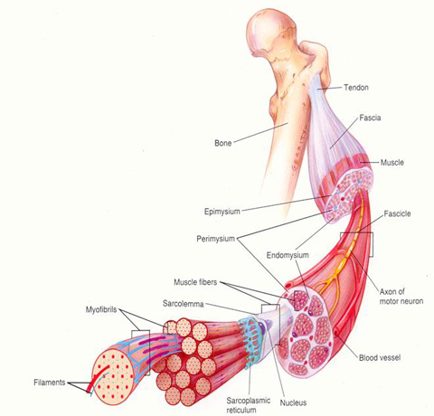
There are over 430 skeletal muscles in the body, and these are made of muscle tissue, connective tissues, nerves and blood vessels. Let’s go over some terminology.
- Epimysium: Also called the deep fascia covers the outside of the muscle and is continuous with the tendon of that muscle.
- Tendon: Thick fibrous connective tissue made of collagen, this is where the muscle attaches to the bone.
- Bone Periosteum: This is a fibrous membrane that covers all bone except for articular surfaces and contains the attachment sites for tendons.
- Proximal: Limb muscles have two attachments to bone, this is one of them it means closer to the trunk.
- Distal: This is another limb muscle attachment and it means farther from the trunk.
- Superior: Closer to the head.
- Inferior: Closer to the feet.
- Origin: The attachment site of the muscle that is more proximal.
- Insertion: The attachment site of the muscle that is more distal.
- Muscle Fibers: Muscle cells can run the entire length of the muscle and are about as thick as a human hair (50 to 100 micrometers), they are multinucleated and have a striated appearance when viewed under magnification.
- Fasiculi: Is a group of muscle fibers under the epimysium, and can contain up to 150 fibers.
- Perimysium: A connective tissue that surrounds the fasiculi, a deeper layer of mysium basically.
- Endomysium: The deepest mysium, as it were.
- Sarcolemma: Surrounds the individual muscle fibers and is a membrane which contains calcium stores which are important for allowing the muscle to contract.
- Motor Neuron: Also called a motor nerve is what carries the electrical signal from the spine to the muscle in order to contract.
- Neuromuscular Junction: Where the motor neuron meets with the muscle fiber.
These structures are parts of the macro and micro structure of the skeletal muscle. One more note about the connective tissues: the epimysium, perimysium and endomyium are all continuous with the tendon, and when the muscle is stretched this tension is transferred to the tendon, which is useful for force production from elastic recoil.
A motor neuron innervates or connects to many muscle fibers, sometimes as many as several hundred. The grouping of a motor neuron to the muscle fibers it innervates is known as a motor unit. One interesting feature of this arrangement is that when a motor neuron sends a signal to contract, all muscle fibers it innervates will contract, no ifs ands or buts about it. But we have many motor units, the more of them we turn on, the more muscle area we activate.

Photo Source: http://fau.pearlashes.com/anatomy/Chapter%2014/Chapter%2014_files/AP_I_7.jpg
Above is a picture of one muscle fiber, there are many components to this structure. Notice that the entire muscle fiber is surrounded by the sarcolemma, which is surrounded by the endomysium (not pictured). The sarcoplasm which is the cytoplasm of the muscle cell, (depicted in blue) is a fluid substance that contains contractile components which can be seen in the diagram as round bundles called myofibrils which consist of myosin and actin filaments. In the sarcoplasm we can also find fats, glycogen, enzymes, mitochondria, other proteins and of course water. The myosin and actin filaments also called thick and thin filaments, make up the functional contractile component of the muscle. This happens through the formation of cross bridges, which is the linkage of a myosin head to an actin binding site. This happens in sections known as sarcomeres, see image below.

Photo Source: http://www.ucl.ac.uk/~sjjgsca/MuscleSarcomere.gif
The thin filaments are wound together in two strands like a double helix. The thick filaments are joined to adjacent thick filaments by the M bridge in the H zone. Actin filaments are joined together with neighboring actin at the Z line. This pattern is continuous throughout the entire myofibril and gives us continuity in the larger picture of the skeletal muscle as a whole. Six actin filaments surround each myosin filament, and each actin filament is surrounded by 3 myosin filaments. All of these factors that affect the arrangement of the muscle give the striated appearance but especially the presence of the Z line.
The A band is made of the myosin filaments and is the darkest section, the I band is the space on either side of the A band and contains the actin filaments and Z lines as well. The H zone is the middle of the sarcomere which is made up of just the myosin filament. Surrounding each myofibril is what is known as the sarcoplasmic reticulum, which stores calcium ions. Coming off of the sarcoplasmic reticulum are transverse tubules or T-tubules which run perpendicular to the myofibrils and go around and in between them inside the muscle fiber. This is the way that electrical signals are conducted to the muscle and allows a signal to reach the entire depth of the muscle almost simultaneously. A grouping of two sarcoplasmic reticulum vesicles and a T-tubule is known as a triad. So how does all of this end in a muscle contraction? It is known as the sliding filament theory.
Sliding Filament Theory
Basically, the myosin filaments pull on the actin filaments and bring the Z lines closer together, and the H zone and I bands shrink. But remember, the A band will remain the same, because the length of the myosin filaments is not changing. In this way the sarcomere shrinks and thus the entire muscle shortens. Now, there are two other proteins on actin that I have not mentioned yet which make this shortening possible, troponin and tropomyosin. Tropomyosin blocks actins binding site for myosin. When calcium is released however (from the sarcoplasmic reticulum) it floods the cytoplasm of the muscle cell and binds to the TNC subunit of another protein called troponin. Troponin then pulls on tropomyosin and uncovers the binding sites allowing cross bridge formation to happen and the muscle to contract. So, without calcium present in the muscle cell, we cannot contract our muscle. What releases the calcium is the electrical signal that travels down the sarcolemma and into the T-tubule, the myosin head is ready to create a cross bridge as soon as it is given the opportunity. This reaction is powered by the breakdown of ATP into ADP and AMP which cocks the myosin head and pulls on the actin creating a muscle contraction. It actually takes more energy to break the cross bridge and disengage, and this will keep happening as long as tropomyosin is out of the way.
Neuromuscular System
Motor neurons have connections to muscle fibers and this creates a motor unit, there are different types of motor units with some having many innervations (many muscle fibers for the same motor neuron) for powerful movements and some that have much fewer such as the eyes and hands which allows us to have fine motor control. The all or none principle states that all muscle fibers that are part of a motor unit will contract when the motor unit tells them to, there are no partial contractions. This brief contraction is called a twitch, and many twitches must summate in order to produce noticeable force. This is because the signal to contract will release calcium and allow cross bridge formation, but if more signals to contract are not sent, the calcium is pumped out of the cell, and the cross bridges are broken before maximal force can be produced. Decreasing time between twitches will result in greater summation of forces and power output. This is until tetanus, which is the maximal amount of force a muscle fiber can produce.
Muscle Fiber Types
There are three types of skeletal muscle fibers, type I, type IIa and type IIb or IIx as it is now called. These fibers differ in the rates at which they can develop a twitch, as the name might suggest. They also have differences in the amount of ATPase enzyme, aerobic and anaerobic capacity and so on. Generally type I fibers are slow twitch, they are very efficient and resistant to fatigue but do not produce as much force as type II or fast twitch fibers. These fibers are less efficient, tire quickly, but produce much greater force and more quickly. Type IIa is more aerobically efficient than type IIb (x) but both are less so than type I.
Motor Unit Recruitment Patterns During Exercise
The simplest way to look at this is that we recruit muscles from slow to fast twitch, or from low threshold motor units to high. In this way we ensure we can use our body to produce coordinated movements and save energy and prevent muscle burnout for simple activities like writing and brushing our teeth. Then as demand increases, higher threshold motor units are recruited when we need speed and power.
Preloading
This is an effect that happens when we are using weighted resistance but not isokinetic, hydraulic or friction modulated resistance. When overcoming the inertia of a resistance from weight, the muscle has time to fully activate and get the maximum number of cross bridges formed and thus we can generate more force in the early ranges of motion.
Proprioception
Proprioceptors are specialized sensory receptors inside your joints, muscles and tendons. They give us proprioception which is our ability to judge where our body is in space relative to each of our body segments. Muscle spindles are one such receptor inside our muscles which are innervated by gamma motor nerves and are intrafusal. When our muscle is put into a loaded stretch the muscle spindle will send a signal back to the spinal cord and send an excitatory signal back to the same muscle fiber. This is important for injury prevention when we receive impact or slip and fall, the muscle quickly contracts under a fast or powerful stretch in order to stop the muscle fibers from being ripped apart. Golgi tendon organs do basically the opposite job, and they are located in the tendons as the name would suggest. When the muscle is contracting this pulls on the tendon which stretches the GTO which sends a signal back to the spinal cord and an inhibitory inter-neuron and back to the same muscle fiber. This prevents us from using maximal force which could protect us from ripping our tendons off our bones. Weight training has been shown in research to decrease the GTO excitability and raise this threshold.
Cardiovascular System
The heart is essentially two pumps made of atria and ventricles, atria feed the ventricles and ventricles feed the lungs or body. The right side of the heart goes to the lungs (pulmonary circulation) the left side goes to the body (peripheral circulation). We also have valves, of which there are two types, AV valves and semilunar valves. The valves are passive and stop blood from moving backwards through the circulatory system. The right AV valve is called the tricuspid valve, the left AV valve is called the mitral valve. The semilunar valves are known as the aortic and pulmonary valves and both have three triangular flaps that create one solid seal when closed (during diastole). The heart has its own conduction system made of pacemakers and conductive material to propagate these signals. The key features are the sinoatrial node, atrioventrical node, atrioventricular bundle, left and right bundle branches, and purkinje fibers. The heart is self-pacing due to the nature of the myocardium which is self-excitatory. The SA is normally the driving force behind each heart beat since it has the fastest intrinsic rate of depolarization. Next in line would be the AV node and then the walls of the ventricles themselves. The discharge pattern and propagation of the electrical signal through the conductive pathways of the heart allows the heart to beat as a syncytium and effectively pump blood through the entire body.
Electrocardiogram (ECG or EKG)
An ECG gives us a visual representation of the heart and tells us what is going on both physically and electrically with the heart. There are associative norms for the distances and amplitudes of every single segment on an ECG, this is an extremely useful tool for diagnostic testing and disease detection. The P wave represents atrial depolarization, the QRS complex ventricular depolarization, and the T wave ventricular repolarization.

Photo Source: http://www.meditech.cn/images/ecg-info/figure4.jpg
Blood Vessels
In a nutshell we have an arterial system and a venous system, arteries carry blood away from the heart and veins towards it. Arteries and veins both are surrounded by smooth muscles which allows our autonomic nervous system the ability to dilate and constrict them to redirect blood flow as needed. Arteries can be constricted so much as to cut blood flow off to an area entirely, and veins can dilate to act as a blood storage reservoir. Capillaries are intermediaries where gasses and other substances are exchanged from tissues to blood, they are very small and thin, about as wide as a single red blood cell.
Respiratory System (last section!)
As far as this system goes what we are really concerned with is the exchange of carbon dioxide and oxygen. It starts in the nasal passages, which aren’t just for our sense of smell, they also warm, moisten and purify the air for our lungs. The trachea is considered the first generation respiratory passage, and the bronchi the second. By the time you get through all the bronchioles and to the alveoli, we have 23 generations of respiratory passages, that’s a lot of little branches. We control this air moving in and out both by using our diaphragm and expanding our rib cage using the intercostal muscles. During rapid breathing such as in exercise the abdominal muscles also aid in breathing. Thanks to pressure gradients, we only use 3% to 5% of our energy on breathing at rest and 8% to 15% during exercise. Concentration gradients allow materials to diffuse from blood to tissues or vice versa in the capillary beds, molecules simply move from high concentration to low concentration, making this process completely automated and no energy is needed for chemical reactions.
Reference:
Baechle, Thomas R. Roger, Earle W. (2008). 3rd Edition. ESSENTIALS of STRENGTH
TRAINING and CONDITIONING. National Strength And Conditioning Association.


Leave a comment